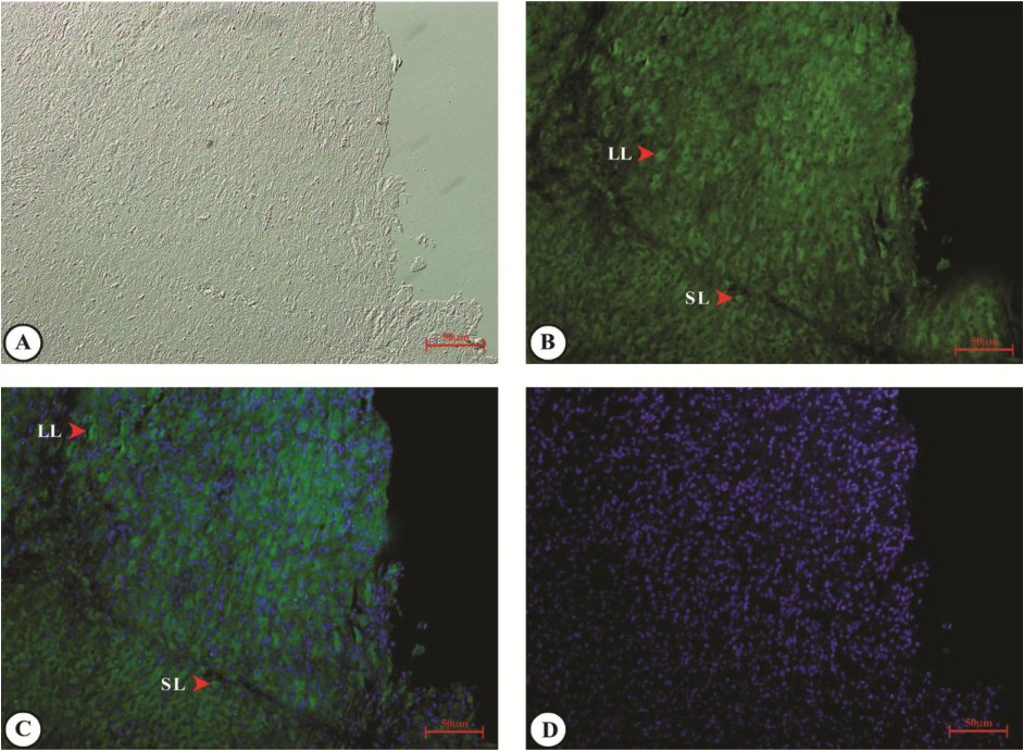
Fig. 5. Immunohistochemical localization of CD36 in the late CL stage of riverine buffalo. The 5 μm thick sections of CL were deparaffinised and rehydrated, followed by antigen retrieval. Primary TSP1 antibody was used at 1:200 while the FITC was used at 1:500. DAPI was used to counterstain nucleus. Fluorescent signals were captured by microscopy (Carl Zeiss Micro Imaging GmbH). Representative images from (A) bright field, (B) through (C) indicate intense immunoreactivity in late stages of CL which was localized predominantly in the cytoplasm of luteal cells. No primary antibody was used in the negative control (D). Scale bar = 50 μm. Abbreviations: LL, Large luteal cell; SL, Small luteal cell; FITC, Fluorescein isothiocyanate; DAPI, 40, 6-diamidino-2-phenylindole dihydrochloride.